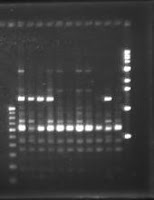
Agarose gel Electrophoresis
Agarose gel electrophoresis?
Gel electrophoresis is a procedure of separation and analysis of macromolecules (DNA,RNA and proteins) and their segments, based on their size and charge. It is used in clinical chemistry, biochemistry and molecular biology for separation of DNA, RNA or protein.
Table of Contents
What is agarose gel electrophoresis?
Gel electrophoresis is a procedure of separation and analysis of macromolecules (DNA,RNA and proteins) and their segments, based on their size and charge. It is used in clinical chemistry, biochemistry and molecular biology for separation of DNA, RNA or protein. Nucleic acids are differentiate by applying an electric field to move the negatively charged molecules through a matrix of agarose or other substances. Shorter molecules move faster and migrate farther than longer ones because shorter molecules migrate more easily through the pores of the gel. Proteins are separated by the charge in agarose because the pores of the gel are too small to sieve proteins. Gel electrophoresis can also be used for the separation of nanoparticles.
Pic: Gel Elecrophoresis.Which reagents are needed for Agarose gel electrophoresis?
- Loading dye (0.25% xylene cyanol, 0.25% bromophenol blue, 30% glycerol and 1 mM EDTA)
- DNA marker (100bp)
Procedure of performing agrose gel electophoresis.
- One μl loading dye was placed on a piece of aluminum foil paper using 0.5-10 μl adjustable micropipette.
- Finally, 3μl extracted DNA was added to it and mixed well using same micropipette.
- The samples were then added slowly to allow them to sink to the bottom of the wells.
- The gel was placed in the gel chamber containing 1XTBE buffer. The final level of buffer was~5mm above the gel. The power supply was then connected and turned on to move the DNA from negative to positive electrode (black to red).
- Electrophoresis was carried out at 120v for about 1.5 hour. After the bromophenol blue dye had reached three-fourths of the gel length, the electrophoresis was stopped and the power supply was automatically disconnected.
Documentation of the DNA samples.
- After electrophoresis, the gel was taken out carefully from the gel chamber and the gel gently washed in running water and placed on a plate which contains Ethidium Bromide (EtBr) solution.
- After staining about 15-20 minutes, the gel was carefully placed on the UV trans-illuminator in the dark chamber of the image documentation system.The UV light of the system was switched on; the image was viewed on the monitor, focused, acquired and saved in a diskette, as well as printed on paper.
Precautions needed to take during test
- As Ethidium Bromide (EtBr) is a powerful mutagen and carcinogen, hand gloves were used when handling anything that had been exposed to Ethidium Bromide.
- A transilluminator produces UV radiation in the 254 nm range. This wavelength can cause eye damage (short term burns, long term cataracts and skin cancers). Thus eye protector should be used while working with it.


Comments
Post a Comment